
Rotary Microtomes & Histopathology Laboratory Equipment Sourcing Guide
Comprehensive technical engineering guide on manual rotary microtomes, semi-automated histocut units, cryostats, automatic tissue processors, and tissue flotation baths for clinical pathology labs, medical colleges, cancer research institutes, and hospital diagnostic centers worldwide.
Quick Answer: What is a Rotary Microtome & What Are Its Core Industrial Specifications?
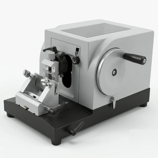
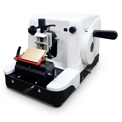
A Rotary Microtome is a high-precision medical laboratory instrument engineered to cut biological specimens embedded in paraffin wax into ultra-thin, uniform microscopic sections typically ranging from 0.5 µm to 60 µm in thickness. Operated either manually via a balanced handwheel or through an automated stepper-motor drive, rotary microtomes utilize vertical mechanical motion translated into micrometer-increment horizontal specimen feed to deliver consistent ribbons for histological examination, cancer biopsies, and anatomical research.
1. The Critical Role of Precision Microtomy in Clinical Diagnostic Pathology
In histopathology, oncology, and anatomical pathology, microscopic diagnosis depends entirely upon the quality of the tissue section placed upon the glass slide. If a tissue slice is uneven, compressed, torn, or irregular in thickness, cellular morphology is distorted, leading to diagnostic delays or misinterpretations. This makes the rotary microtome the foundational workhorse of every pathology department, medical university, and bio-specimen research institute.
As a leading scientific laboratory equipment manufacturer and exporter based in Ambala, India, AJKANT OVERSEAS manufactures a complete ecosystem of ISO 9001:2015 and ISO 13485:2016 certified histopathology equipment. From heavy-base manual rotary microtomes to microprocessor-controlled automated units, rapid cryostats, and automated tissue processors, our equipment is built with German-standard cross-roller bearings and zero-backlash micrometer feed screws to deliver repeatable sub-micron precision under high-throughput laboratory conditions.
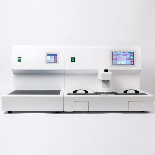
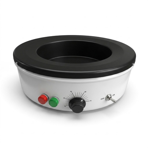
AJKANT Histopathology & Microtomy Equipment Catalog
Directly manufactured by AJKANT OVERSEAS. Click on any model below to inspect detailed factory specifications or request an export quotation.
2. Engineering Anatomy & Working Principle of a Rotary Microtome
The fundamental mechanism of a rotary microtome converts the rotational motion of a side-mounted handwheel into synchronized vertical strokes of a specimen holder, coupled with minute horizontal advancements toward a stationary blade. Here is the mechanical anatomy:
1. Handwheel & Flywheel Assembly
Counterbalanced flywheel design provides frictionless, uniform rotational inertia. Features dual fail-safe lock mechanisms (handwheel lock at 12 o'clock and independent carriage brake) for user safety during blade or block changing.
2. Precision Micrometer Feed Screw
Engineered from case-hardened alloy steel with zero-backlash cross-roller guideways. Translates every rotation into exact 0.5 µm to 60 µm increments without play or thermal drift.
3. Specimen Retraction Mechanism
On the upward return stroke, the specimen chuck automatically pulls back by 5 to 20 µm away from the blade edge, eliminating friction, block heat build-up, and blade dulling.
4. Multi-Axis Specimen Orientation
Calibrated X-Y axis orientation system allows ±8° micro-adjustment and full 360° Z-axis rotation to align the tissue block face perfectly parallel to the knife cutting edge.
3. Complete End-to-End Histopathology Laboratory Workflow & Equipment
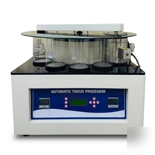
A high-precision microtome works within an interconnected tissue preparation sequence. AJKANT OVERSEAS manufactures the entire equipment chain required to take a raw biopsy specimen from grossing to high-magnification diagnosis:
4. Technical Specifications Comparison Matrix: AJKANT Microtome Models
Compare the mechanical and electronic parameters of our four flagship microtome and cryostat models to select the optimum instrument for your institutional workload:
5. Microtomy Defects: Causes & Corrective Actions Guide
Even with precision microtomes, environmental factors and blade maintenance impact section quality. Below is the clinical troubleshooting matrix utilized by pathology laboratory managers:
Defect: Ribbon Compression & Wrinkling
Symptoms: Sections appear squashed, shorter than the actual block face, and jammed together.
Solution: Warm paraffin blocks must be chilled on ice / cold plate prior to sectioning. Ensure blade clearance angle is between 4° and 7°. Clean wax debris off back of blade holder.
Defect: Chatter & Venetian Blind Striations
Symptoms: Parallel horizontal thick-and-thin ridges running across the section.
Solution: Vibration issue. Verify specimen clamp and blade holder are completely locked. Reduce sectioning speed. Tissue may be over-dehydrated or hard; soften block face with moist gauze.
Defect: Vertical Scratches / Split Ribbons
Symptoms: Long vertical tears or splits dividing the ribbon along the direction of the cut.
Solution: Microscopic nick on the blade edge or hard particulate/calcification in the tissue. Shift disposable blade laterally to a fresh edge. Decalcify tissue blocks if bone/calcium is present.
Defect: Alternate Thick & Thin Sections
Symptoms: One thick slice followed by an extremely thin or skipped section.
Solution: Microtome mechanism play or insufficient blade clearance angle (causing the block face to bounce off the knife facet). Tighten all clamp levers and reset clearance angle to 5°.
6. Why Sourcing Histopathology Equipment from AJKANT Overseas Gives You an Unfair Advantage
Procuring laboratory instrumentation directly from the original manufacturer guarantees engineering integrity, spare parts availability, and factory pricing without middleman markups:
100% In-House Precision Machining
Our manufacturing facility in Ambala, Haryana utilizes computer-controlled CNC lathes and surface grinders to fabricate feed screws and guideways with sub-micron tolerances.
ISO 13485:2016 Certified
Full medical device quality management standard compliance, complete with traceability documentation and factory calibration certificates for every unit shipped.
Global Export-Grade Packaging
Reinforced seaworthy wooden crates with high-density polyurethane foam cradle molds, moisture desiccant bags, and anti-vibration strapping ensure zero damage in international transit.
Turnkey Lab B2B Tenders
We regularly supply government health ministries, university medical departments, forensic science labs, and private hospital networks across the Middle East, Africa, and Asia.
7. Frequently Asked Questions (FAQ) — Histopathology Equipment Procurement
Q1: What is the difference between a Rotary Microtome and a Sledge Microtome?
In a Rotary Microtome, the knife remains stationary while the tissue specimen moves vertically and horizontally on a precision carriage driven by a handwheel, making it ideal for routine paraffin wax sectioning (3–5 µm). In a Sledge Microtome, the tissue block is clamped stationary while a heavy knife slides horizontally across a track, primarily used for cutting very large tissue blocks (whole-organ, neuropathology) or harder materials like bone and industrial plastics.
Q2: What is the ideal section thickness for standard histological routine examination?
For routine diagnostic histopathology with Hematoxylin and Eosin (H&E) staining, sections are universally cut between 3 to 5 microns (µm). For kidney biopsies and lymph nodes, thinner sections of 2 to 3 µm are required to prevent multi-layer cell overlap. For neurohistology, thicker sections of 8 to 15 µm are standard.
Q3: When is a Cryostat Microtome needed instead of a standard Rotary Microtome?
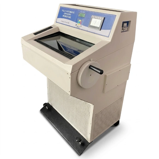
A Cryostat Microtome is essential for intraoperative rapid frozen biopsies during active surgeries (providing results in 10–15 minutes), enzyme histochemistry, and fluorescent immunofluorescence studies where chemical fixation and hot paraffin wax would denature heat-labile enzymes and antigens. The tissue is snap-frozen at -20°C to -35°C and sliced immediately inside the refrigerated chamber.
Q4: What blade types are compatible with AJKANT microtomes?
AJKANT microtomes feature a universal blade holder system compatible with standard high-profile and low-profile disposable microtome blades (such as Feather, Leica, and Sakura standards), as well as conventional reusable steel knives (C-profile wedge, D-profile chisel) via interchangeable clamping inserts.
Q5: Why is specimen retraction important in modern microtomy?
Without specimen retraction, the tissue block brushes against the back facet of the knife during the upward return stroke. This causes friction, builds static charge, generates heat that softens the paraffin, creates vertical micro-scratches on the block face, and rapidly dulls the razor edge. Automatic retraction pulls the specimen back by 15–20 µm during upward travel, tripling blade life and ensuring pristine sections.
Q6: How does AJKANT Overseas package microtomes for sea and air freight exports?
All microtomes and tissue processors are bolted to internal shock-absorbing wooden pallets, vacuum-sealed inside high-barrier moisture protection bags with industrial silica gel desiccants, encased in high-density polyethylene foam cradles, and enclosed in heavy-duty ISPM-15 heat-treated export plywood crates compliant with international customs regulations.
Q7: What quality certifications accompany AJKANT histopathology equipment?
Every batch is manufactured under ISO 9001:2015 quality systems and ISO 13485:2016 medical device standards. Equipment is supplied with CE Declaration of Conformity, individual factory inspection certificates, micrometer calibration sheets, and complete English technical operation manuals.
Equip Your Pathology Laboratory with Direct Factory Sourcing
Whether setting up a new histopathology wing, upgrading to automated microtomy, or bidding on government healthcare tenders, partner directly with AJKANT OVERSEAS for guaranteed precision, factory warranties, and competitive B2B wholesale rates.